Cardiovascular System of the Lower Torso. The posterior wall is next to the perineal body rectum and peritoneal cavity at the pouch of Douglas while the two lateral walls lie against the pelvic diaphragm and major vaginal vessels.

Three Dimensional Posterior View Of The Pelvis Download Scientific Diagram
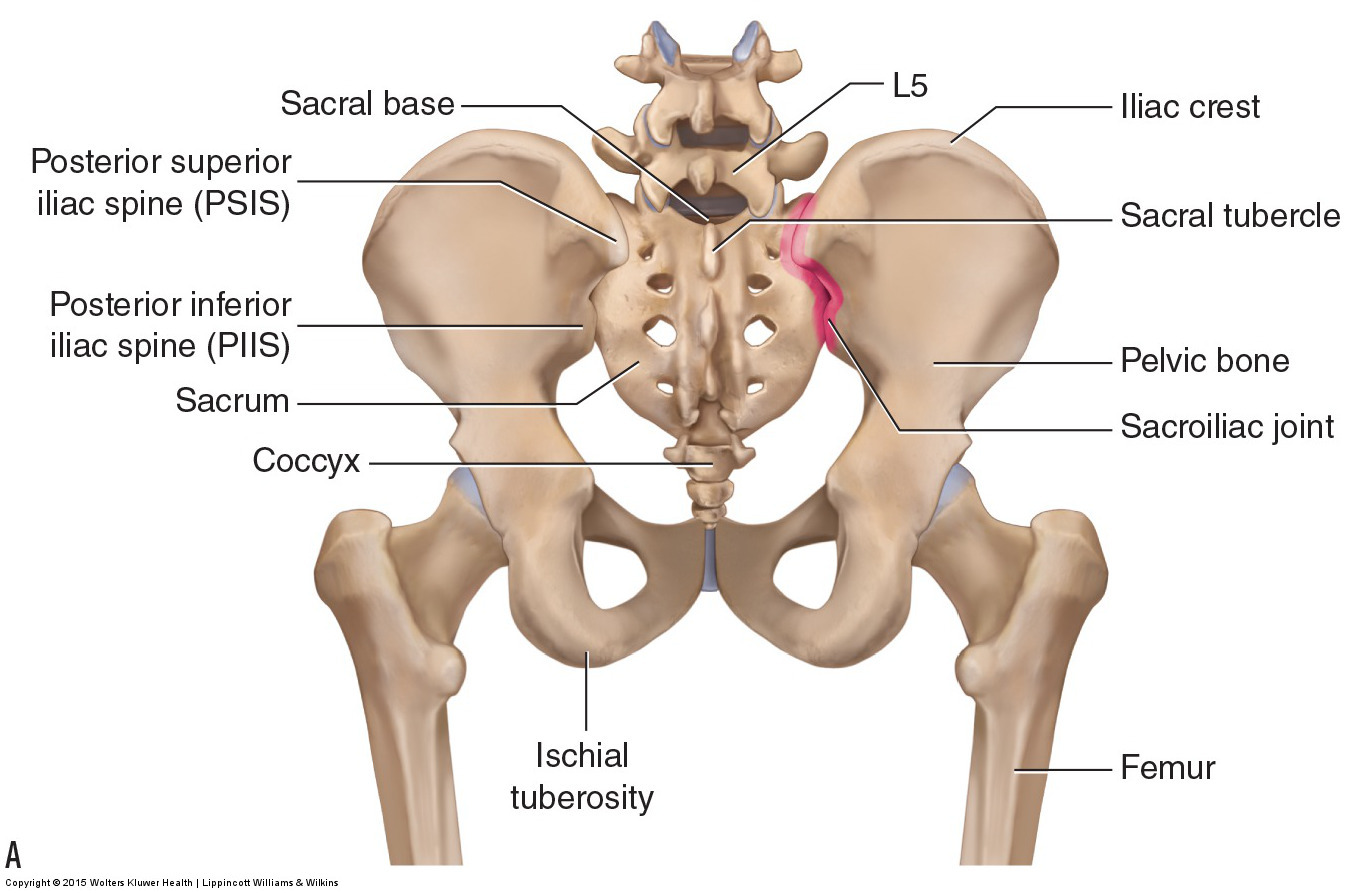
Pelvic anatomy is composed of two innominate coxal bones that articulate with the sacrum and proximal femora.
. Anatomical landmarks within the vagina can be used to locate the position of such structures as the ureter and urethra and warn of their possible involvement in a vaginal laceration. Diagonal conjugate obstetric conjugate etc. Pelvic examinations are common in clinical cases of obstetrics and gynecology and can be performed in various ways ie.
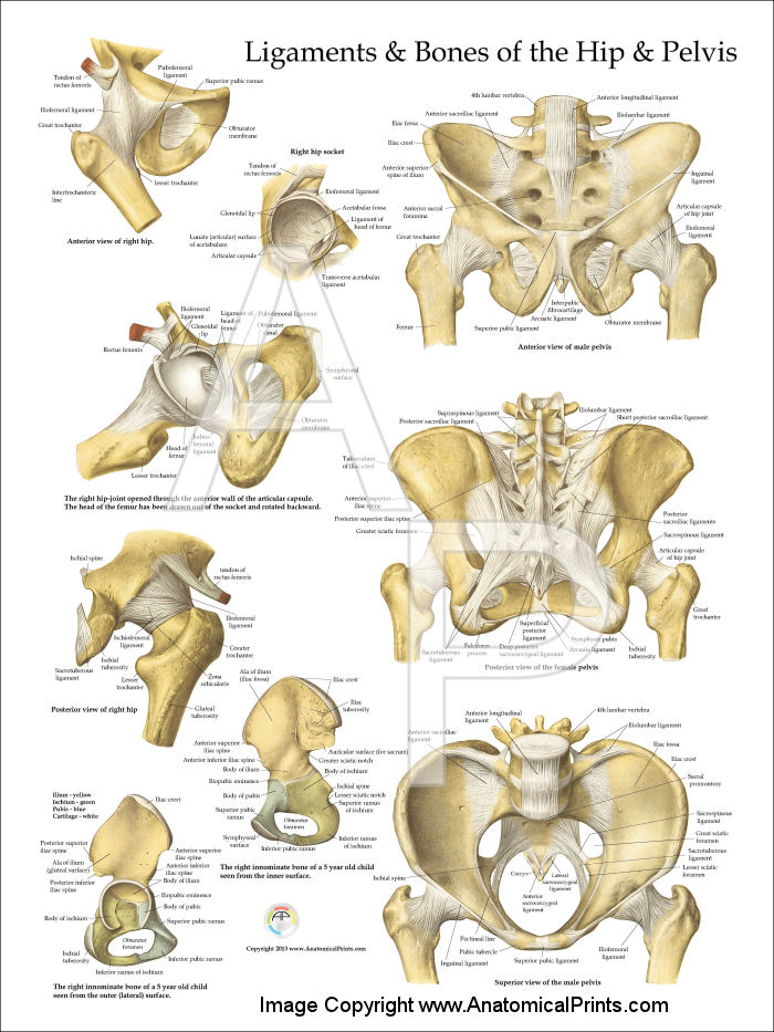
To research radiographic anatomy of the main structure of the pelvic Teepee view including its azimuth direction and view anatomy structure. You may also find sacrospinous ligament lesser sciatic foramen sacrotuberous ligament ischial tuberosity deep posterior. The three bones and three joints composing the pelvic ring have no inherent stability without vital ligamentous structures.
Ilium ischium and pubis meeting in the acetabular fossa at the triradiate fusion. Radiographic Anatomy of the Skeleton. The plane of the pelvic brim faces forward and forms an angle of about 60 degrees to the horizontal.
The female on the other hand has a much wider and more. The parietal pelvic fascia is removed to visualize the embedded autonomic pelvic nerves. Click on a structure to learn its name Show Me a.
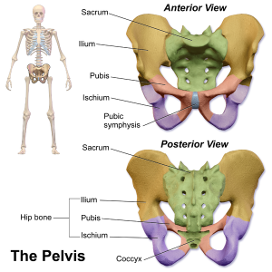
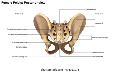
Features that most clearly distinguish the female from the male pelvis include a wider subpubic angle wider sciatic notch and greater distance from pubic symphysis and anterior. The pelvic girdle also known as the hip bone is composed of three fused bones. The pelvis consists of the sacrum the coccyx the ischium the ilium and the pubis.
3D Illustration Of Human Body Skeleton System Pelvis Posterior View Anatomy. The anterior muscles posteriorly tilt the pelvis the posterior muscles anteriorly tilt the pelvis the muscles on the right side elevate the right side of the pelvis and therefore depress the left side of the pelvis and the muscles on the left side elevate the left side of the pelvis and therefore depress the right side of the pelvis. It is usually divided into two separate anatomic regions.
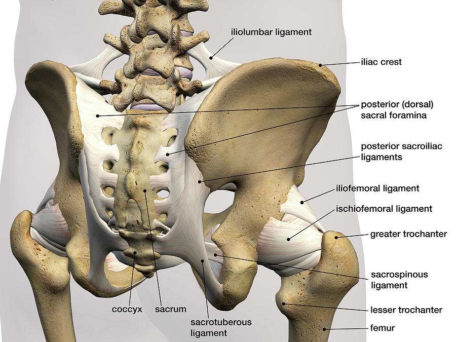
Bones of the Pelvis and Lower Back Posterior View Toggle Anatomy System. Bony pelvis is formed posteriorly by the sacrum and the coccyx and laterally and. We think this is the.
It has fibers that run almost laterally to connect the lateral posterior aspect of the upper two segments to the PSIS and the internal iliac crest. Posterior view - Female Anatomy Muscles. Bony pelvis or pelvic skeleton is formed by hip bones sacrum and coccyx.
Medial view of a right-sided male hemipelvis. Major components of the bony pelvis frontal superior view of the female pelvis. The term pelvis is used to identify the area between the abdomen and the lower extremitiesIt can be divided into the greater pelvis and the lesser pelvis.
Bony pelvis Pelvis ossea The bony pelvis is a complex basin-shaped structure that comprises the skeletal framework of the pelvic region and houses the pelvic organs. We hope this picture Pelvic Region Posterior View can help you study and research. The male pelvis is smaller and narrower with a thinner pubic symphysis.
Download Human Skeleton System Pelvis Anatomy Posterior View Stock Illustration and explore similar illustrations at Adobe Stock. The ilium ischium and. The posterior section is located in the deep depression between the ala of the sacrum and the PSIS.
In this image you will find the posterior superior iliac spine iliac crest tubercle of the iliac crest anterior superior iliac spine greater sciatic foramen the acetabular margin in it. The pelvic girdle and pelvic spine. Topographic anatomy of the posterior pelvic compartment.
Although conditions are uncommon pelvis-based dislocations hernias and prolapses are present in a dynamic range of patient populations1 Responsible for supporting upper body weight the. A The posterior pelvic compartment is delimited from the urogenital compartment by the rectoprostatic septum Denonvilliers fascia. Furthermore 11 investigators reviewed identified abstracts and selected those reporting on posterior female pelvic and vulvar anatomy for full-text review.
Digestive System of the Lower Torso. The sacrum and two innominate bones. The pelvis is a ring structure made up of three bones.
Each innominate bone is composed of three united bones. From June 2013 to June 2014 adult pelvic CT examination results were filtered excluding skeletal deformities and pelvic osseous destruction caused by tumors trauma etc. Most of which reflect the role of childbirth in the female.
The male pelvis is different from a. This online quiz is called Posterior view of Pelvic Anatomy SI ligaments. For more anatomy content please follow us and visit our website.
The pelvic region is the area between the trunk or main body and the lower extremities or legs. Pelvis -- AnteroPosterior AP View Unlabelled. Download scientific diagram Anatomy of the pelvis.
The structure of the pelvis supports the contents of the abdomen while also helping to transfer the weight from the spine to the lower limbs. We are pleased to provide you with the picture named Pelvic Region Posterior View. Human Body Skeleton System Pelvis Posterior View Anatomy.
From inception of the study to April 6 2018 MEDLINE database was used to search for 40 terms relevant to the posterior female pelvis and vulvar anatomy. The pelviss frame is made up of the bones of the pelvis which connect the axial skeleton to the femurs and therefore acts in weight bearing of the upper body. Bone And Ligaments Of Pelvis Posterior View.
The data of 20 mm contiguous CT scan. Radiographic Anatomy of the Skeleton. The pelvis is the lower portion of the trunk located between the abdomen and the lower limbs.
Muscle Diagram Black Man Male Body Names. Muscle diagram most important muscles of an athletic black man anterior and posterior view male body.
Pelvis And Hip Anatomy Poster
Rear View Of Male Pelvis Hip Leg Photograph By Hank Grebe

Pelvis Anatomy Recon Orthobullets
Ilium Physiopedia

The Pelvic Girdle And Pelvis Anatomy And Physiology I
Muscles Of The Pelvis
Bones Of The Lumbar Spine And Pelvis
Skeleton Pelvis Posterior View 3d Illustration Stock Illustration 474011278
0 comments
Post a Comment